What is organ-on-a-chip technology?

Request Information
Organ-on-a-chip is a technology combining cells and tissues grown inside microfluidic chips with the aim of recapitulating physiological processes of human organs. The applications of organ-on-a-chip range from disease modeling and drug testing to personalized medicine and the development of cell and gene therapies. There are a diverse array of organ-on-a-chip devices available and Axion’s innovative platforms can noninvasively monitor cells in microfluidic systems allowing the study of complex physiological processes.
Fast, flexible, organ-on-a-chip imaging
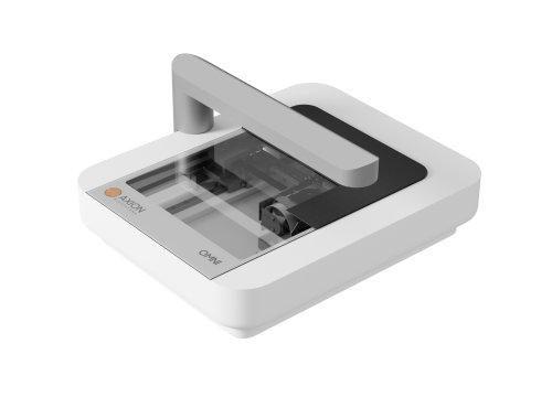
Microfluidic and organ-on-a-chip technology has advanced rapidly with the development of custom and commercial devices. The versatility of the Omni platform is ideal for imaging-compatible devices.
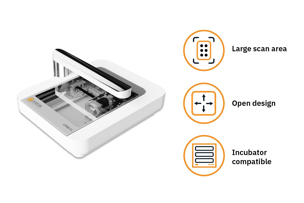
Advantages:
- Large scan area – compatible with any device layout
- Flat open design – doesn’t interfere with connecting tubes or cables
- Incubator compatible – keep cells in optimal conditions during your experiment
Organ-on-a-chip applications
-
Track 3D spheroid formation in microfluidic cell culture>
-
Investigate chemotaxis in microfluidic devices>
-
Model neuronal subtype interactions>
-
Isolate and measure neurite activity>
Track 3D spheroid formation in microfluidic cell culture
>
Purpose: To study self-assembly of individual spheroids in a microfluidic chip under perfusion.
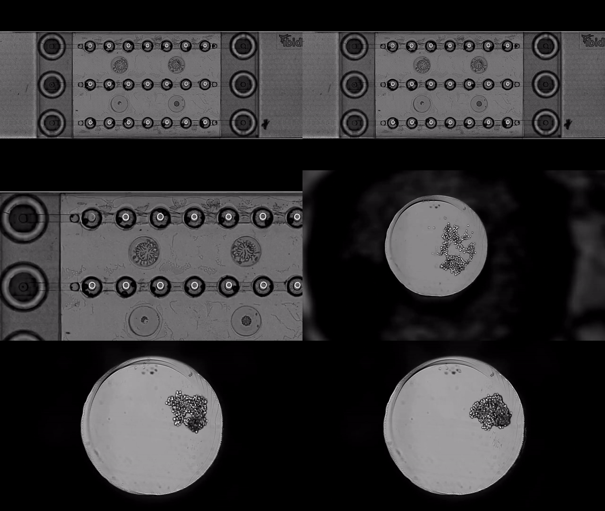
HeLa cells cultured in a μ-Slide Spheroid Perfusion (ibidi, 80350) were monitored using the Omni platform.
Result: HeLa cells started to aggregate and self-assemble into spheroids during the first 20 hours post-culturing. Perfusion in a microfluidic device can ensures optimal nutrition during long-term spheroid cultures.
Investigate chemotaxis in microfluidic devices
>
Purpose: To examine chemotactic migration of human neutrophils through a microscopic maze. Neutrophil chemotaxis is a critical immune response to detect and move toward damaged tissue or infections.
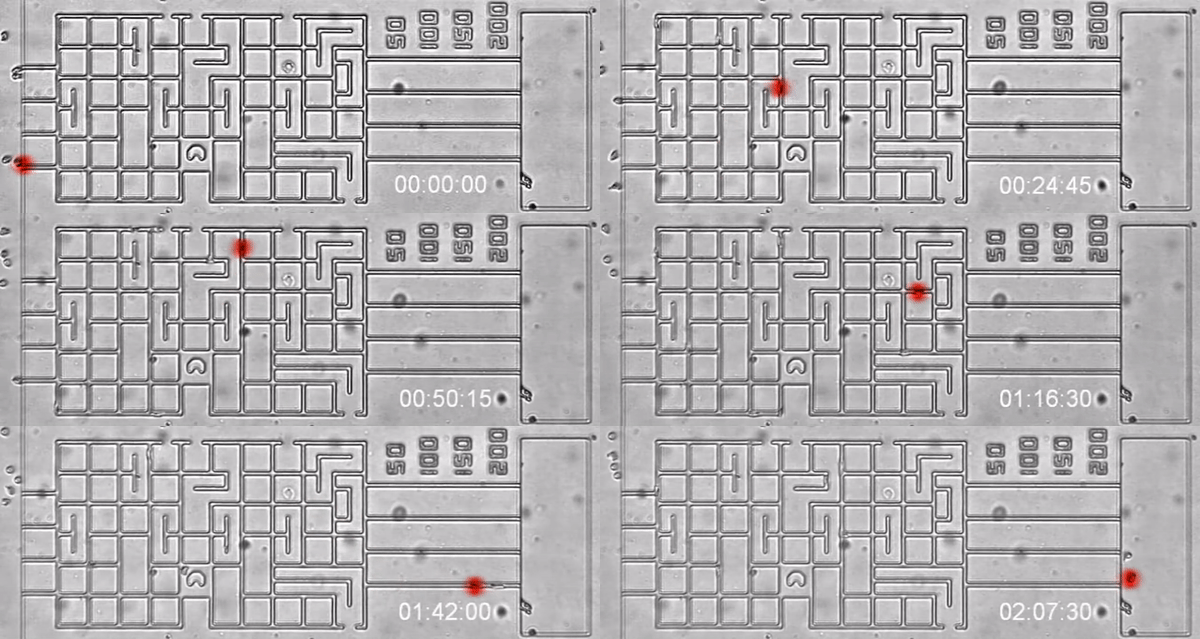
Human neutrophils were added to the microfluidic maze containing LTB4 chemoattractant. The cells were imaged using the Lux platform.
Result: The neutrophils migrated through the microscopic channels towards the LTB4 chemoattractant.
Model neuronal subtype interactions
>
Purpose: To demonstrate synaptic connectivity between different neuronal subtypes. Brain-on-a-chip models can shed light on the interaction between different brain regions.
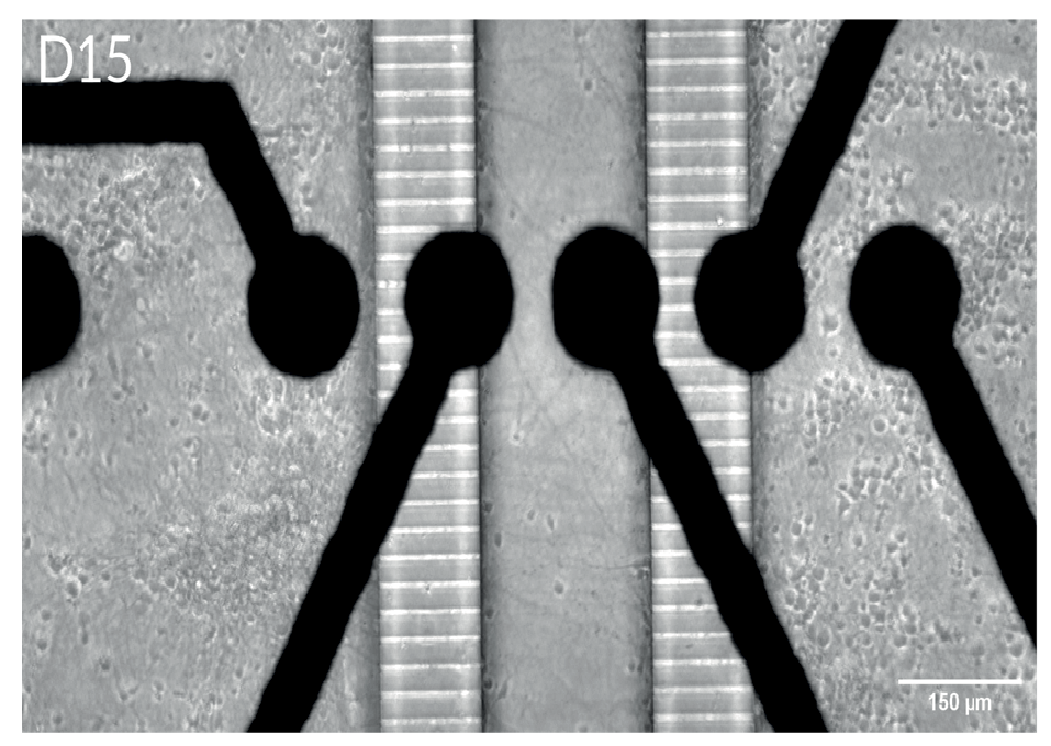
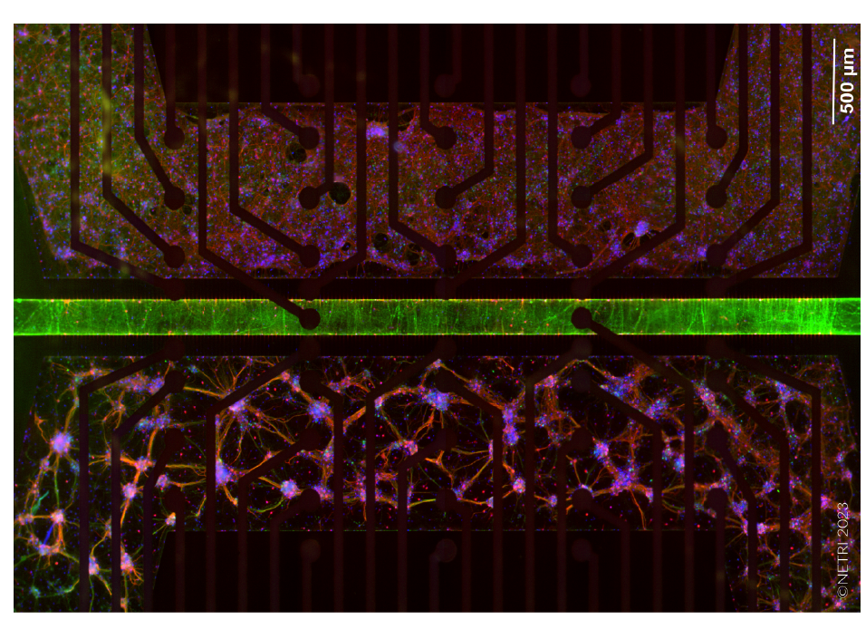
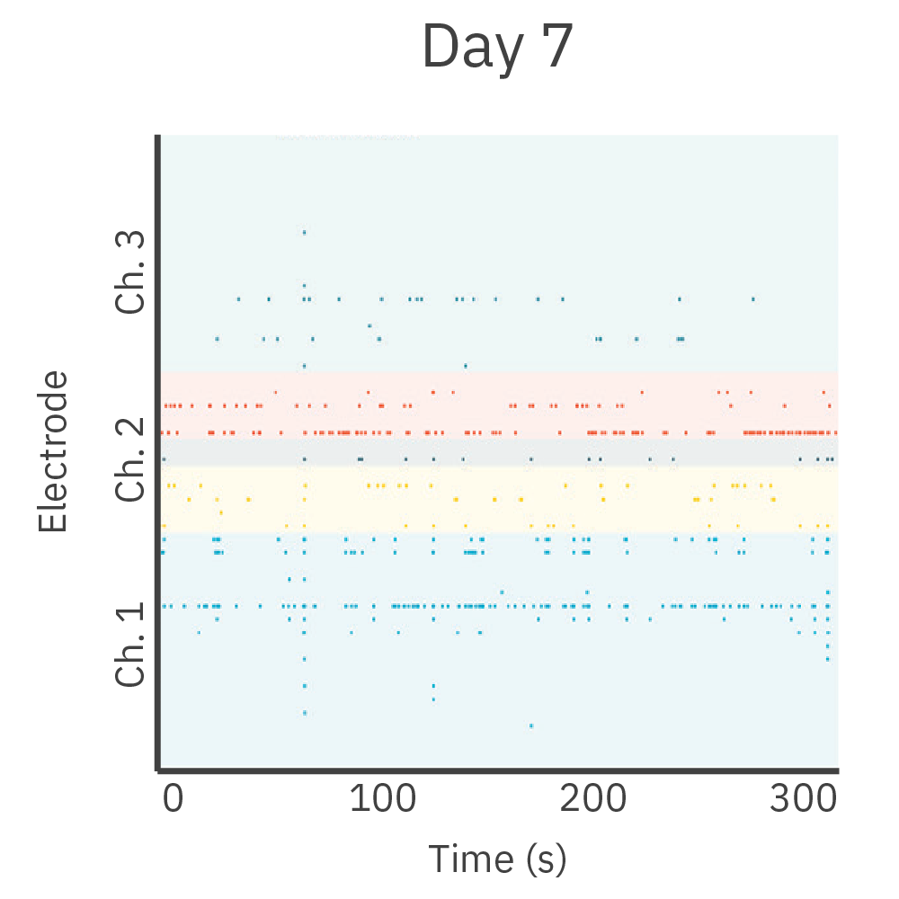
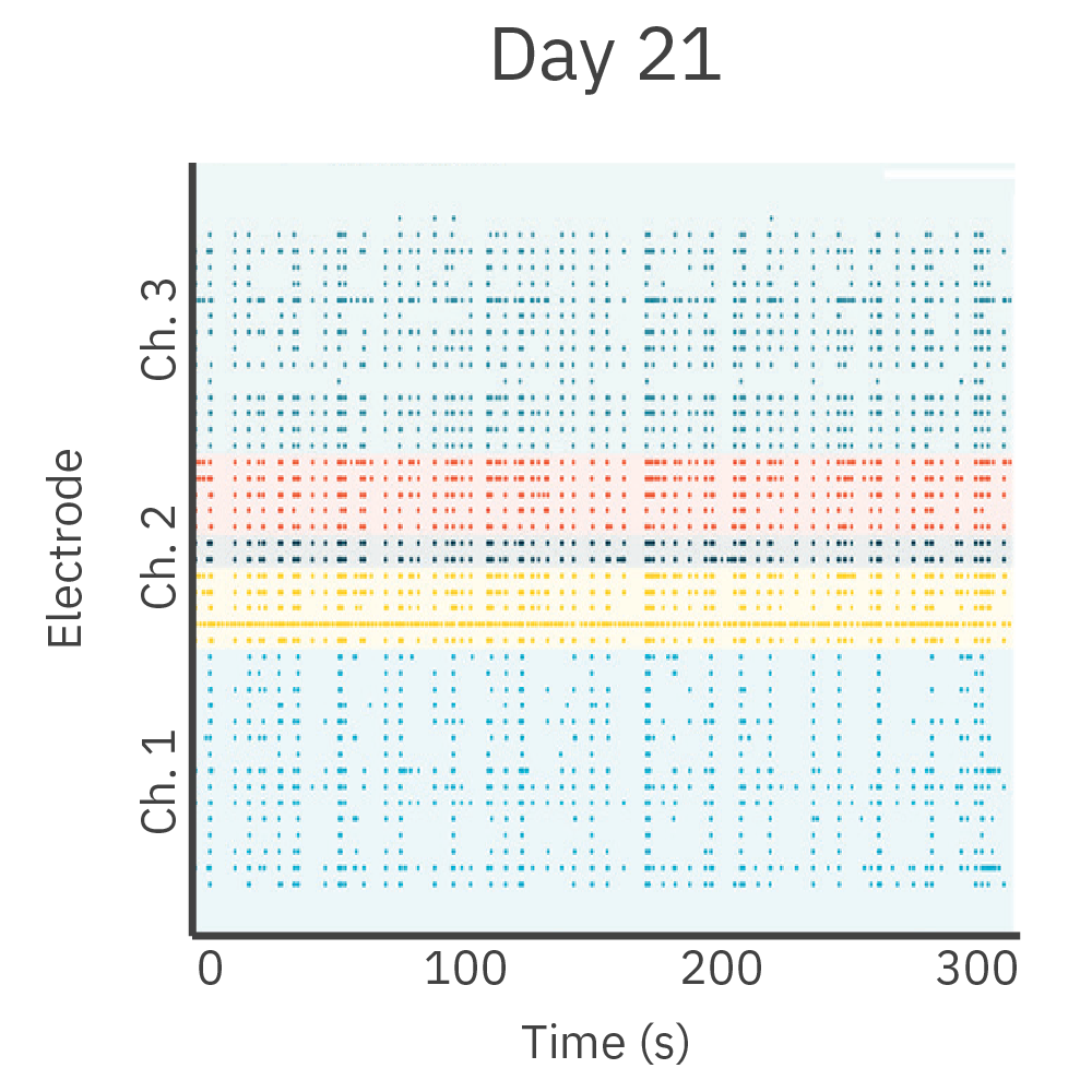
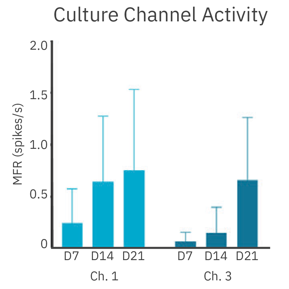
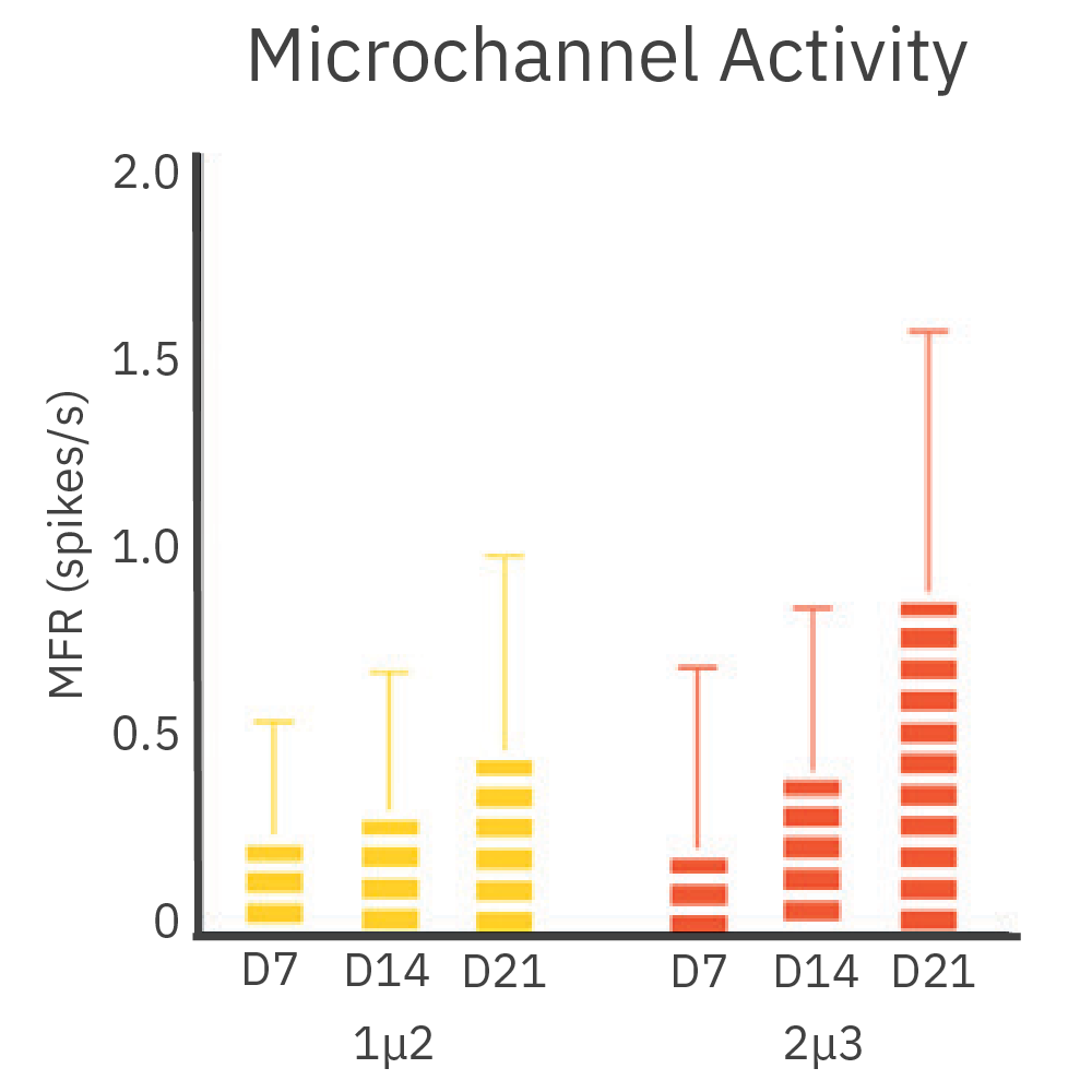
Cortical and hippocampal neurons were cultured in separate chambers of the DuaLink MEA (Netri) and recorded on the Maestro MEA platform over 21 days.
Results: Cultures increased synchronous firing between chambers as new synapses formed over time.
Isolate and measure neurite activity
>
Purpose: To monitor growth and activity in neurites. Understanding the growth and function of neural projections could aid in the development neuroregenerative medicine.
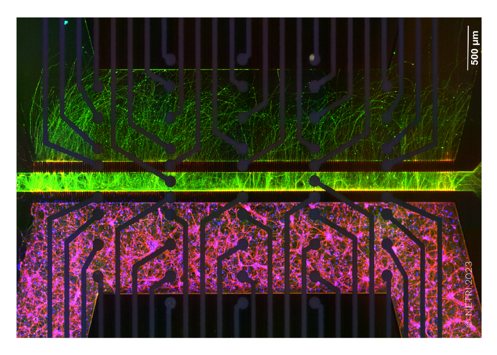
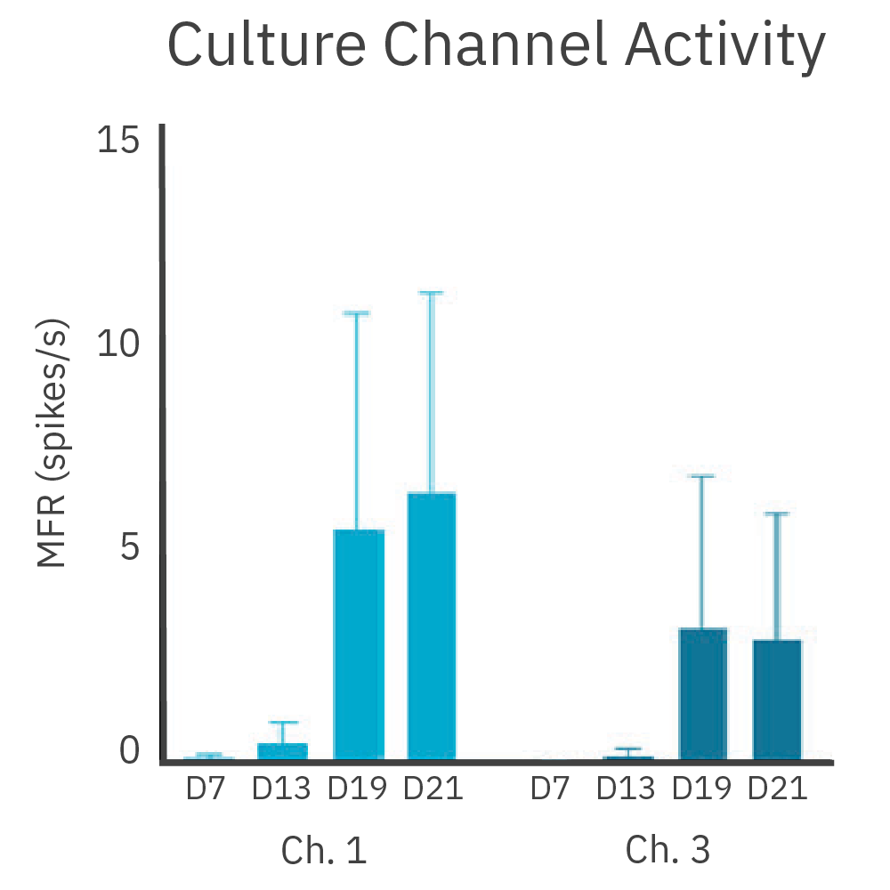
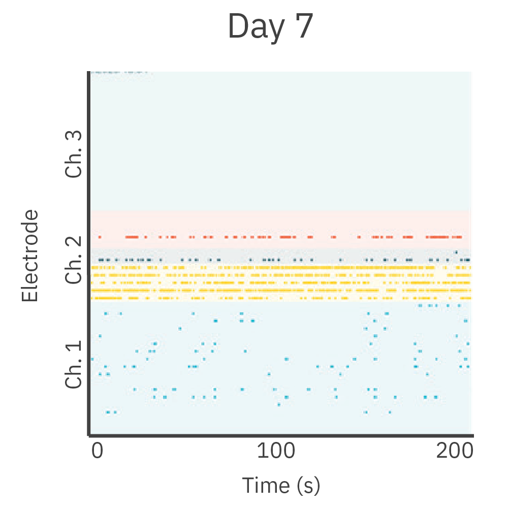
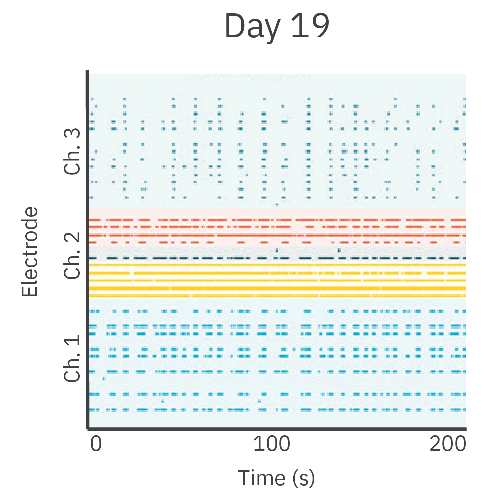
Cortical neurons were cultured in one chamber of the DuaLink MEA (Netri) and recorded on the Maestro MEA platform.
Results: As new neurite projections covered the empty chamber, increasing activity was observed. This activity was synchronous to the activity in the cortical chamber.

Featured Content: Organ-on-a-chip Publications
>> Human Spinal Organoid-on-a-Chip to Model Nociceptive Circuitry for Pain Therapeutics Discovery
>> Osteogenesis and osteoclastogenesis on a chip: Engineering a self-assembling 3D coculture
FAQ:
Organ-on-a-chip technology refers to microscale systems that contain tissues and cells grown inside microfluidic chips, mimicking the organ’s functionality and microenvironment. Organ-on-a-chip platforms represent a combination of microfluidics and tissue engineering, offering unique insights into human biology.
What are the benefits of using organ-on-a-chip models?
The aim of organ-on-a-chip technology is to more accurately replicate human physiological responses, minimizing the need for animal testing. Cells are cultured and subjected to controlled conditions, allowing the study of disease mechanisms and drug responses with high precision.
Why should I use the Omni platform with my organ-on-a-chip device?
For organ-on-a-chip devices that are imaging compatible, the Omni offers a large, flat scan area with open sides to run any additional cables or tubing. This makes the system compatible with a wide variety of devices. Scanning can be automated inside the incubator, minimizing the disruption of the device and its controlled environment.
What research areas and applications can benefit the most from organ-on-a-chip technology?
Organ-on-a-chip technology has applications in drug development, toxicity testing, personalized medicine, organ/disease modelling, and regenerative medicine.
What types of organs or tissues can be replicated using organ-on-a-chip technology?
Organ-on-a-chip technology allows researchers to replicate various organs and tissues, including brain, kidney, lung, heart, liver, and more.
What are the advantages of neural organ-on-a-chip/brain-on-a-chip models
Neuronal cell bodies are primarily located in the brain and spinal cord, connecting to other cells (skin, neurons, muscle, etc.) by their axonal projections. The compartmentalization of microfluidic devices allows researchers to study neural innervation and interaction with the different cell types with high precision.
Are there any Maestro MEA-compatible neural organ-on-a-chip devices available?
Yes. The Neurofluidics microfluidics chips by Netri is compatible with the Maestro MEA platform.