Fluorescence Module
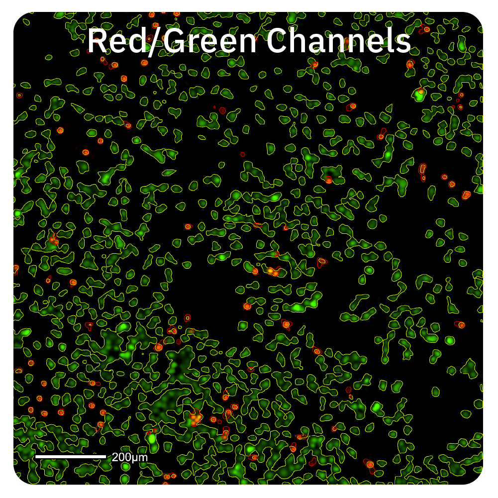
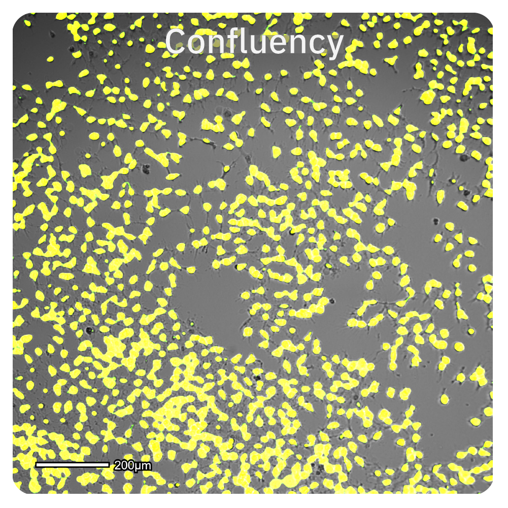
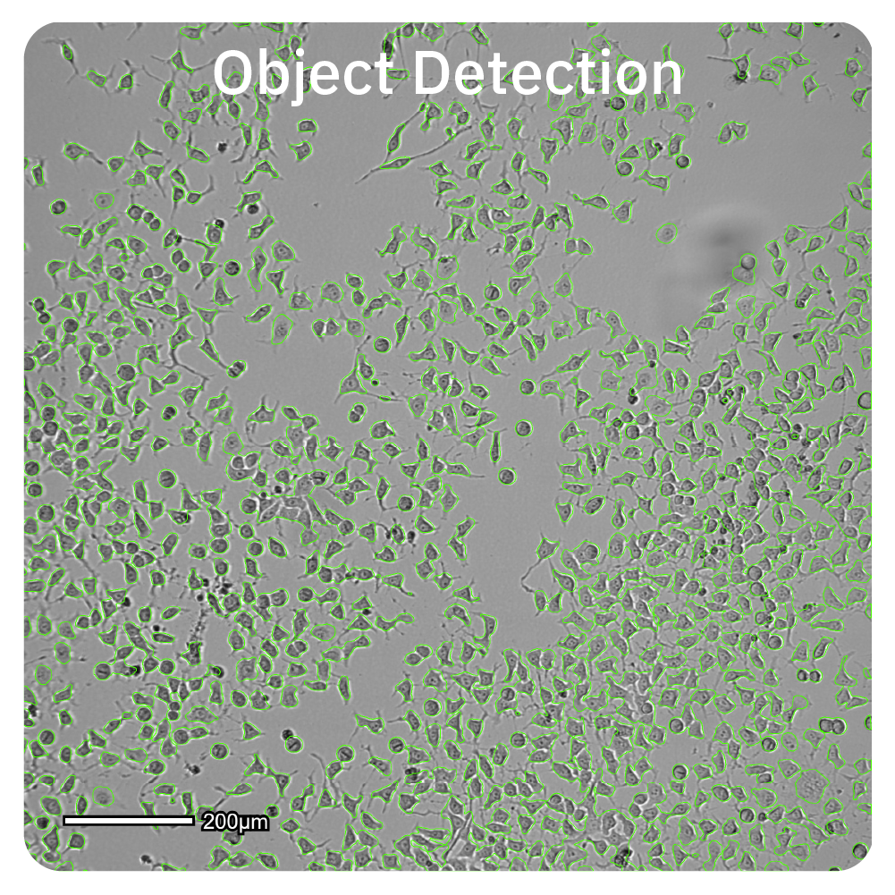
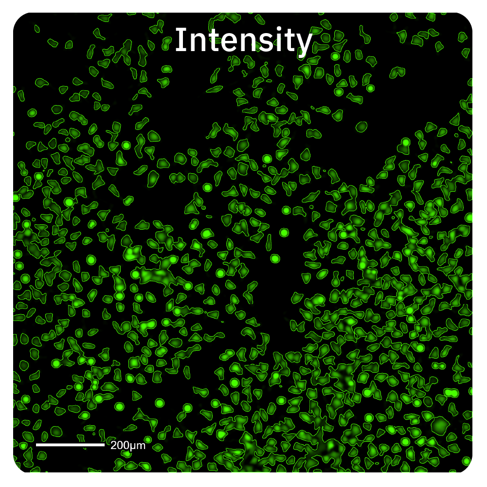
The Fluorescence Module expands the Omni™ and Lux™ live-cell imaging functionality for fluorescence-based assays. Equipped with green and red fluorescence channels for measuring fluorescence confluency, intensity, and object count, this module is a versatile tool designed to analyze cell cultures, straight from the incubator.
Key Features
Add fluorescent assay capabilities with confluency, intensity, and object detection
- >> Versatile fluorescence assays — Assay complex biology in real-time – adding fluorescence capabilities to your Lux or Omni.
- >> Efficient data visualization — Review multiple snapshots and quickly navigate through images with FL map view.
- >> Simple fluorescence acquisition — Don’t waste time at the microscope. Quickly set up the Omni to automatically image your cultures while your cells remain in the incubator.
- >> Automated analysis — Access results instantly to evaluate your cultures and generate publication-ready graphs.
Overview
Study your cells using fluorescence live-cell assays
Fluorescence live-cell imaging reveals kinetic cellular processes over time. Quantify fluorescent tags and reporters, and track cellular processes behind proliferation, cytotoxicity, transfection, and organization of 3D cell cultures with the Fluorescence Module.
Assaying kinetic cell killing
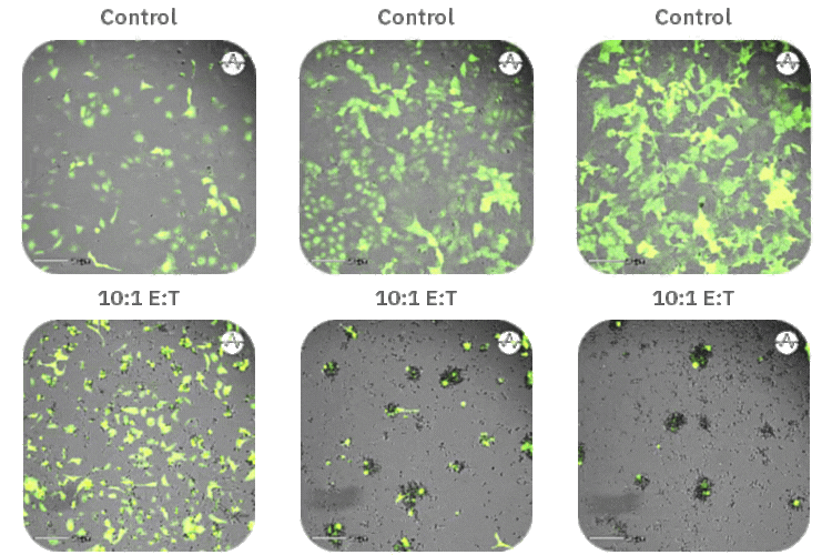
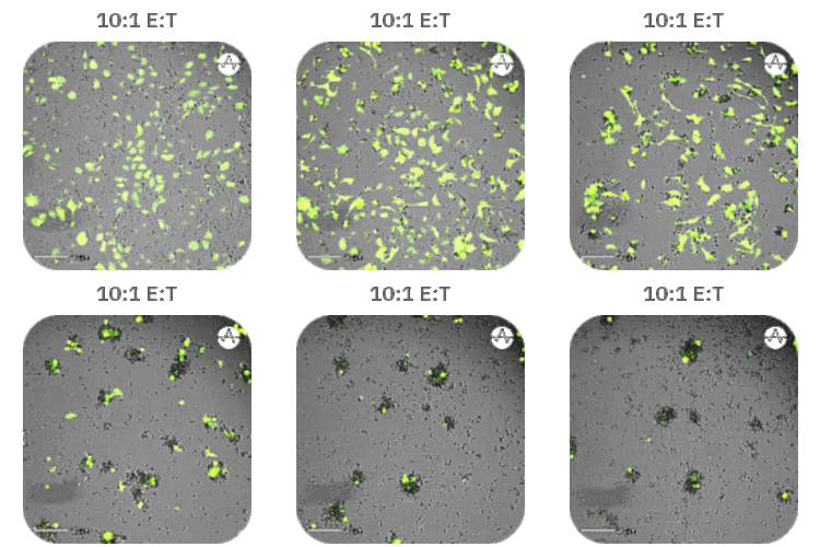
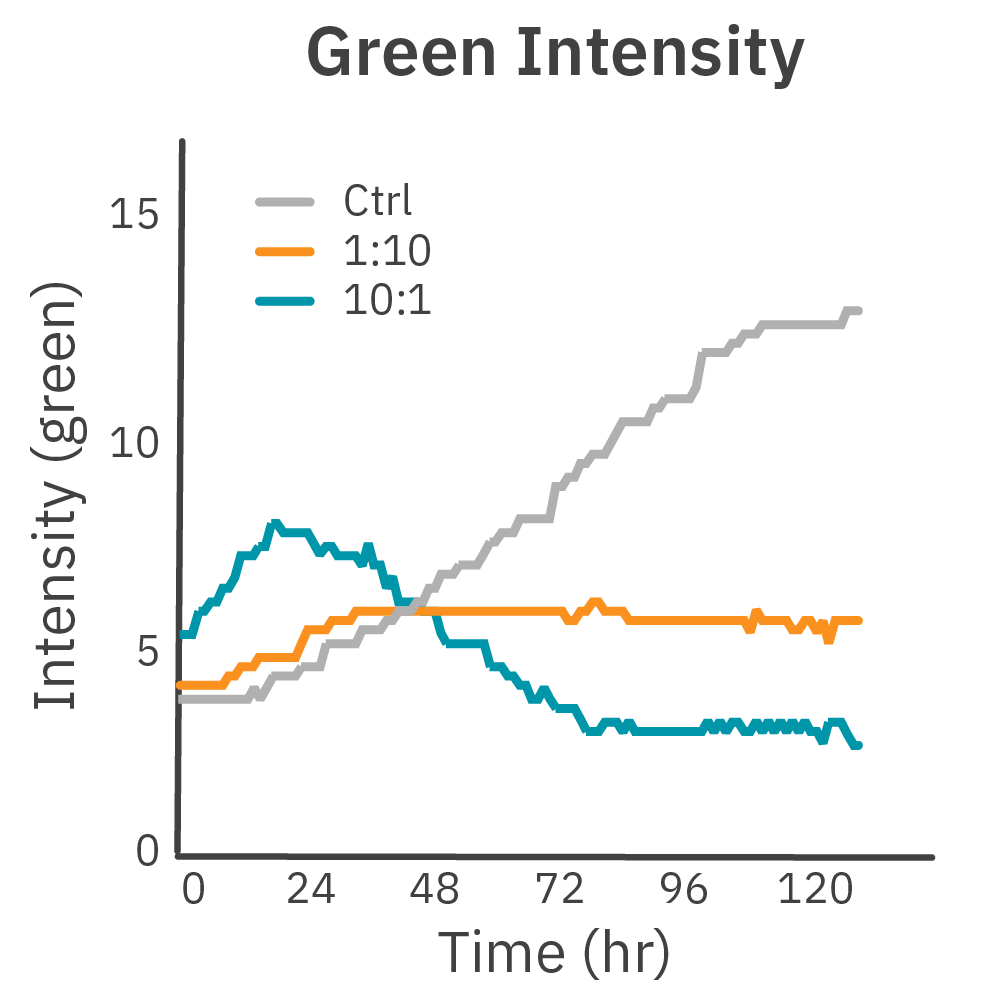
Immune cell-mediated killing of GFP-labeled cancer cells by CAR T cells is quantified over time by fluorescent intensity showing a dose-dependent response.
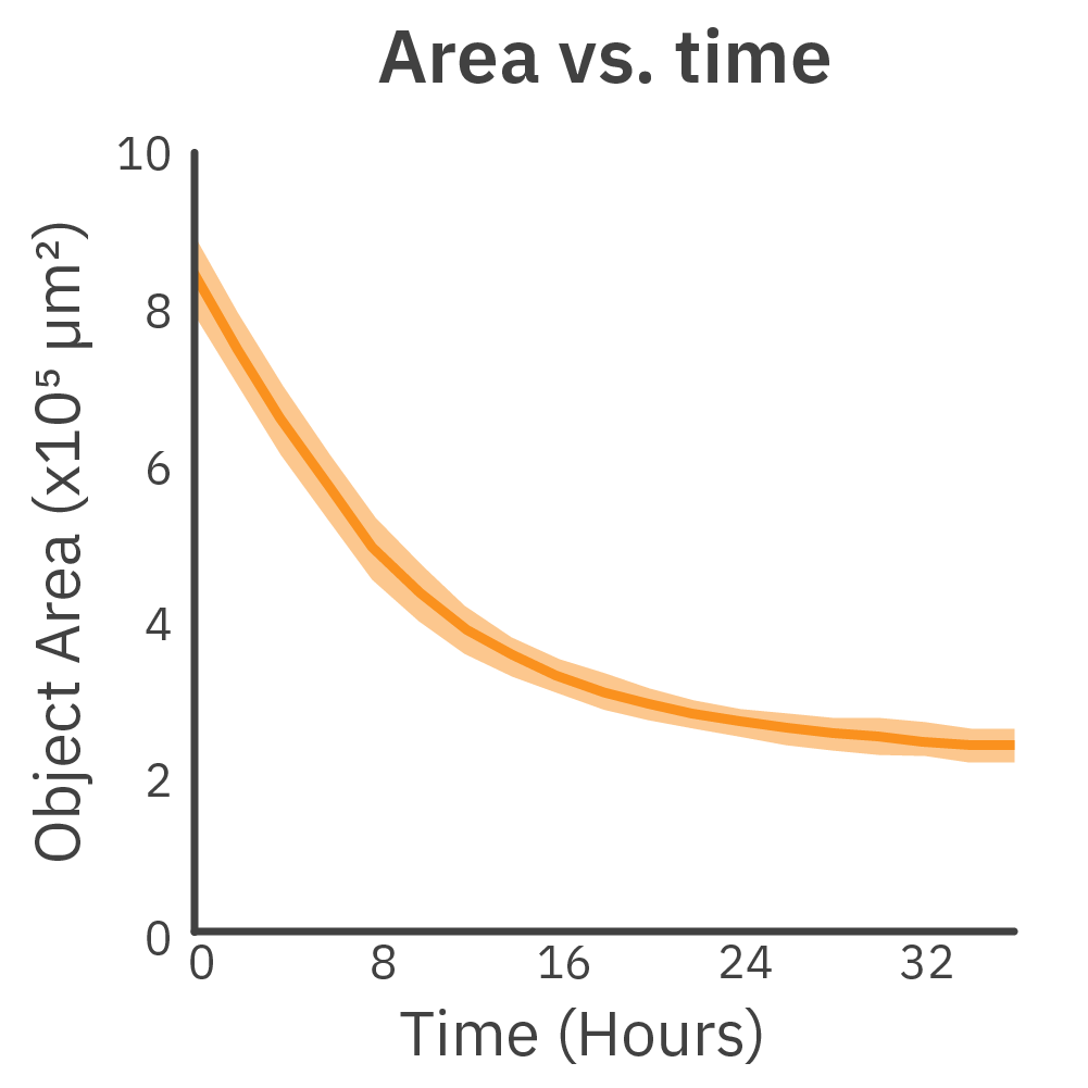
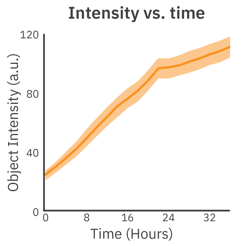
Measuring fluorescence of 3D cultures
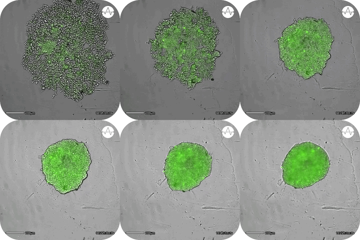
Real-time imaging tracks the formation of spheroids in GFP-labeled cells. The size (area) and fluorescent intensity of the spheroid are measured as it forms.
Quantify and track real-time metrics automatically
The Fluorescence Module can quantify:
- >> Confluency (%)
- >> Intensity
- >> Object count
- >> Object area (µm2)
- >> Object intensity
- >> Average object intensity
