Your neural culture looks healthy. But is it functioning as a network?
Morphology and molecular markers can provide valuable information about cell identity and condition, but they do not directly show how neurons fire, communicate, or organize into functional networks. To understand what a neural culture is actually doing, researchers need a functional measurement.
Microelectrode array (MEA) technology provides this functional readout by recording extracellular electrical activity noninvasively across many locations in a cell culture at once. Because the cells remain intact, researchers can repeat measurements over time and use multiwell MEA systems to compare cultures and experimental conditions in parallel.
In this video, see how microelectrode arrays work, how MEA recordings capture neural activity, and how researchers can use firing patterns and network behavior to characterize electrically active cell cultures over time.
What will you learn?
- What microelectrode array (MEA) technology is and how it works
- How MEA electrodes detect extracellular electrical activity from neurons
- How raster plots visualize neural activity across an electrode array
- What firing, bursting, synchrony, and network oscillations reveal about a neural culture
- How noninvasive, multiwell MEA recordings support longitudinal studies across multiple conditions
What is a microelectrode array?
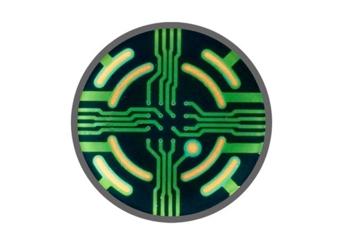
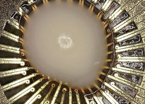
A microelectrode array (MEA), also known as a multielectrode array, is a grid of small electrodes used to measure electrical activity from cells.
When the electrodes are embedded in the culture surface, researchers can plate electrically active cells directly over the array using standard cell culture techniques. The electrodes then detect changes in extracellular voltage associated with cellular electrical activity without disrupting the cells.
This provides an alternative to techniques such as manual patch clamp, which require specialized training and typically measure individual cells one at a time. MEA technology instead enables researchers to simultaneously monitor electrical activity across many cells and locations within a culture.
How does MEA technology measure neural activity?
Many cell types in the human body are electrically active, including neurons, cardiomyocytes, retinal cells, and muscle cells. In neural cultures, MEA recordings can provide insight into both individual neuronal activity and the behavior of the broader network.
As neurons develop in culture, they extend processes, form synaptic connections, and create spontaneously active networks. When a neuron near an MEA electrode fires an action potential, the resulting change in extracellular voltage can be detected by the electrode as a spike.
Multiple electrodes simultaneously record these signals across the culture. Instead of observing activity from only one neuron or location, researchers can therefore investigate how electrical activity develops and propagates throughout a neural network.
How is neural network activity visualized?
One way to visualize MEA recordings is with a raster plot.
On a raster plot, each tick mark represents the time at which a spike was detected, while each row represents activity recorded by an individual electrode. Combining recordings from the electrodes across a well creates a visual representation of neural activity throughout the culture.
Researchers can use these recordings to investigate questions such as:
- How active is the neural culture?
- Do neurons fire randomly or in organized patterns?
- Does activity occur in bursts?
- How long do bursts last?
- How coordinated is activity across the network?
These patterns provide information about the functional behavior of the neural culture that goes beyond simply determining whether individual neurons are electrically active.
Characterizing neural network behavior with MEA
MEA recordings can be quantified using metrics that describe different aspects of neural network activity, including overall activity, bursting behavior, synchrony, and network oscillations.
Together, these measurements create an activity profile of the culture.
This profile can act as a functional fingerprint. Changes in the profile may reflect differences in:
- Cell composition
- Culture health
- Maturity
- Genetic background or modification
- Chemical or compound effects
Because MEA measurements capture functional activity, researchers can compare these profiles between cultures, experimental conditions, or timepoints.
Why use MEA for longitudinal neural recordings?
One advantage of MEA technology is that recordings are noninvasive. Cells do not need to be fixed, labeled, or destroyed to collect an electrical activity measurement.
Because the cells remain intact, researchers can return to the same culture repeatedly and observe how its functional activity changes over time.
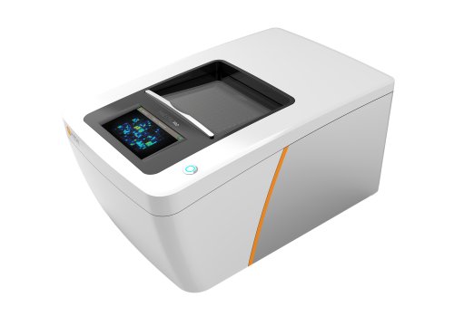
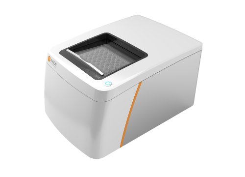
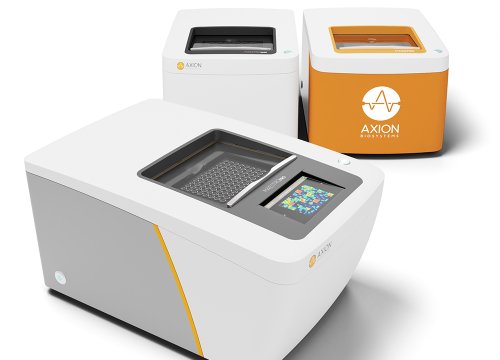
With multiwell MEA systems, multiple cultures and experimental conditions can also be measured in parallel. The Maestro multiwell MEA platform, for example, can monitor neural activity from cultures across up to 96 wells, enabling longitudinal experiments that extend over weeks or months.
Repeated measurements can be particularly useful when the biological question involves changes in neural function rather than a single endpoint.
What can researchers study with MEA technology?
MEA provides functional electrophysiological data that can be applied across a variety of cell-based research questions.
Researchers can use MEA recordings to:
- Characterize iPSC-derived neural cultures
- Follow neural network development and maturation
- Investigate functional changes associated with disease
- Compare genetically modified and control cultures
- Measure cellular responses to compounds
- Screen treatments for safety or efficacy
Because the same cultures can be measured repeatedly, researchers can examine how these functional phenotypes emerge and change throughout an experiment.
From electrical signals to functional insight
Understanding cellular function is critical for studying how biological models develop, respond to treatment, and change in disease.
Microelectrode array technology makes it possible to measure electrical activity across a cell culture without disrupting the cells. For neural models, individual spikes, network bursts, synchrony, and oscillatory behavior provide complementary information about how neurons communicate as a functional network.
By capturing these signals across multiple electrodes and over repeated measurements, MEA assays provide a window into the functional behavior of neural cultures over time.
Watch the video to learn how microelectrode array technology captures neural activity and provides functional insights into electrically active cell cultures.
For research use only. Not for use in diagnostic procedures.