Product Videos
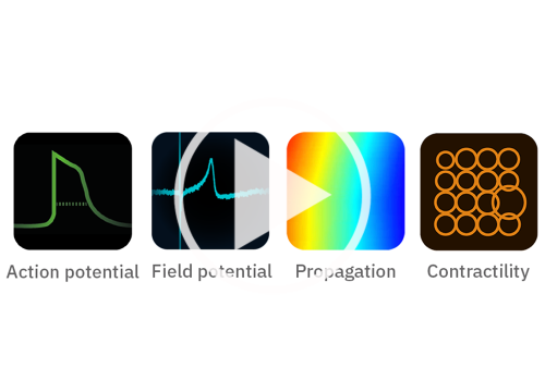
1 system, 4 cardiac assays
Author:
Axion BioSystems
Product:
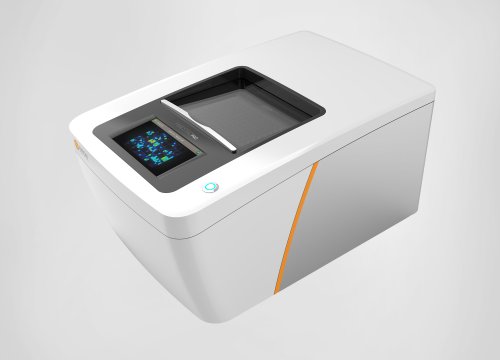
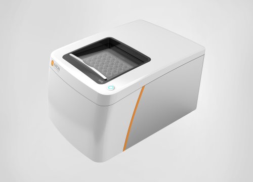
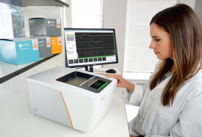
Maestro Pro,
心筋モジュール,
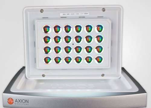
CytoView MEA プレート,
Maestro Edge,
BioCircuit MEA プレート,